The Nobel Prize for Chemistry rewards researchers for major advances in studying the infinitesimal bits of material that are the building blocks of life.
Recent prizes have gone to scientists who developed molecular “machines” with controllable motions and who mapped how cells repair damaged DNA, leading to improved cancer treatments.
The 2017 prize, worth 9 million kronor ($1.1 million), is being announced on October 4 by the Royal Swedish Academy of Sciences.
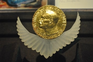
Jacques Dubochet, Joachim Frank and Richard Henderson won the 2017 Nobel Prize in Chemistry for developing cryo-electron microscopy, which simplifies and improves the imaging of biomolecules.
This method has moved biochemistry into a new era.
Researchers can now freeze biomolecules mid-movement and visualise processes they have never previously seen, which is decisive for both the basic understanding of life's chemistry and for the development of pharmaceuticals.
Chemistry is the third of this year’s Nobel Prizes
The medicine prize went to three Americans studying circadian rhythms - Jeffrey C. Hall, Michael Rosbash and Michael W. Young.
The physics prize went to Rainer Weiss, Barry Barish and Kip Thorne for detecting gravitational waves.
The literature winner will be named on October 5 and the peace prize will be announced on October 6.
Moving Past Electron Microscopy: Know More- For many years - in the 1970s, the electron microscope was the only way to look into the cell and observe the minute beings that play such an important role in our lives such as viruses.
- However, the powerful beam of the electron microscope would destroy biological material, so it was believed that such microscopy could only reveal images of dead cells and dead organisms.
- Also it was then impossible to view solutions as water would evaporate under the microscope’s vacuum.
- That was until this year’s laureate Richard Henderson came on to the scene.
- Finally, in 1990, 15 years after he had published the first model, Prof. Henderson achieved his goal and was able to present a structure of bacteriorhodopsin at atomic resolution.
Cryo Electron Microscopy- “Cryo”, short for cryogenic refers to very low temperatures.
- Though the actual temperature is not well defined, it is below minus 150°C.
- In the context of electron microscopy, it refers to the fact that the object to be imaged is frozen to such low temperatures to facilitate being studied under the beam of the electron microscope.
- This method is so effective that even in recent times, it has been used to image the elusive Zika virus: When researchers began to suspect that the Zika virus was causing the epidemic of brain-damaged newborns in Brazil, they turned to cryo-EM to visualise the virus.
- Over a few months, three dimensional (3D) images of the virus at atomic resolution were generated and researchers could start searching for potential targets for pharmaceuticals.
Scientific Contributions in This Field- Prof. Frank had long worked to find a solution to just that problem.
- In 1975, he presented a theoretical strategy where the apparently minimal information found in the electron microscope’s two-dimensional images could be merged to generate a high-resolution, three-dimensional whole.
- Between 1975 and 1986, Prof. Frank succeeded in merging two fuzzy images of a molecule to get a three-dimensional image.
- In 1978, Prof. Dubochet was recruited to the European Molecular Biology Laboratory in Heidelberg to solve another of the electron microscope’s basic problems: how biological samples dry out and are damaged when exposed to a vacuum.
- The solution he envisaged was to freeze water rapidly so that instead of solidifying into a crystalline solid, it freezes into a disordered state, which is like a glass.
- Though a glass appears to be solid, it is actually what is called a supercooled liquid in which individual molecules are arranged at random instead of a periodic crystalline solid structure.
Prof. Dubochet realised that if he could freeze the water to form a glassy state, what is known as vitrified water, it would not dry up when excited by the beam. - In the early 1980s, Prof. Dubochet cooled water so rapidly that it solidified in its liquid form around a biological sample, allowing the biomolecules to retain their natural shape even in a vacuum.
- In 1984, he published the first images of a number of different viruses, round and hexagonal, that are shown in sharp contrast against the background of vitrified water.


